Tandimplantater – Guided Implant
Metoder og risici
Traditionel metode
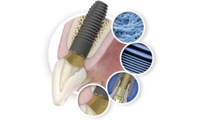
Der er naturligvis altid fordele og ulemper ved tandimplantater. Fordelene taler for sig selv, men det er en omstændig proces, der kræver tid, og typisk tager det op til 6 måneder for implantatet og kæbeknoglen at vokse sammen. Denne sammenvoksning kaldes osseointegration. Læs mere >her<
Med traditionel metode indsættes implantat ved en operation, hvor hele området frilægges. Derefter sys området sammen, og der afventes opheling af implantat typisk ½ år, førend aftryk til abutment og krone tages og fremstilles. Helingsprocessen påvirkes imidlertid negativt af tobak, dårlig mundhygiejne, ukontrolleret diabetes, stråle-/ kemo-/ steroid- terapi, hvorfor behandling med implantat frarådes i disse tilfælde.
Indsættelse af tandimplantater foretages som en kirurgisk operation, og en kirurgisk operation vil altid være forbundet med en række generelle risici. Disse generelle risici omfatter infektion, mulige skader på nervevæv og kraftig blødning under eller efter operativt indgreb.
Med digital 3D scanning af tandsæt og kæber kan sandsynlighed for risici nedsættes.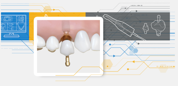
Med digital 3D scanning af tandsæt og kæber kan planlægning visualiseres virtuelt i 3D og reducere omfang af operation.Implantat, abutment og temporær krone bliver special fremstillet efter den foretagne 3Dscanning med 3Shape scanner og 3D CBCT.
Princippet giver mulighed for kort efter operation at indsætte midlertidig krone (3D printet krone), således at der ikke er en „tom plads‟ i tandrækken. Efter ½ år fremstilles permanent krone (MK krone).
Princippet med minimal operation har altså den fordel, at indgrebet er minimeret. Opheling af tandkød og indheling af implantat er derfor fremskyndet, men der skal fortsat udvises forsigtighed og respekt om det krævende miljø, som mundhulen altid vil være, lige gyldigt hvor godt forberedt implantatet indsættes. Derfor anbefales lige efter operation med indsættelse af implantat, at der indsættes healing abutment (se nedenfor), som er designet specifikt efter den aktuelle scanning af tandkød i området.
Guided Implant
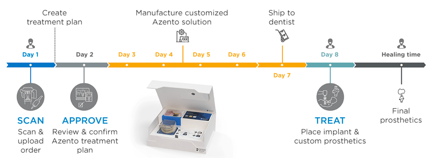
Den ovennævnte metode kaldes Guided Implant, og jeg bruger Azento systemet med de gennemprøvede implantater fra DentsPly Sirona ASTRA Implant System. Jeg har brugt ASTRAs implantater siden 1992.
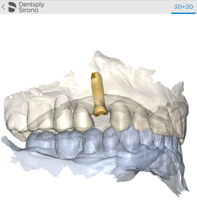
Kæbeknogle (de indre konturer) 3Dscannes med CBCT scanner, og tandsæt (de ydre konturer) scannes med digital 3D scanner. Begge dele sker i klinik på Ulrikkenborg Plads. Sammensættes (stitching (1)) de to 3D scanningstyper i 3D, kan den helt nøjagtige position af implantat vælges.
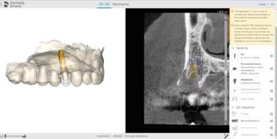
Med Implant Planner 3D software kan operation visualiseres og planlægges, og implantat, abutment og temporær krone special fremstilles custom made. Metoden er præcis og reducerer omfanget af kirurgi.
Abutment
Healing og permanent
Abutment kaldes forbindelses delen mellem selve implantatet og tandkronen. Abutment fastsættes på implantat med en skrue.
Abutment systemet anvendt i forbindelse med Azento Guided Implant/ASTRA Implant System er navngivet Atlantis.
Udformning mod tandkød er med Azento Guided Implant custom made. Dvs. at abutment er special fremstillet efter 3D scanning. Hermed fremstilles abutment nøjagtigt efter tandkødets overflade det pågældende sted, hvor implantat planlægges indsat.
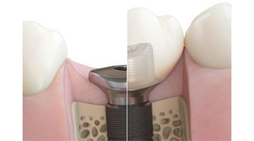
Viser situationen under indsættelse af implantat, at det ville være klogt at afvente en ophelingsperiode, indsættes hygiejnisk ophelings abutment – healing abutment (se foto ovenfor). Er vurderingen, at der kan indsættes permanent abutment + krone, indsættes i stedet dette. En af mange fordele ved Azento Guided Implant er, at vælges en ophelingsperiode med healing med healing abutment er udformning mod tandkød nøjagtig ens med det permanente abutment, hvorved der opnås en harmonisk opheling af tandkødet, og udformning af tandkød efter opheling passer nøjagtig til det permante abutments facon, der mod tandkødet er identisk med healing abutment.

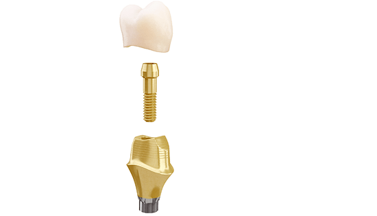
Abutment er special fræset fra en blok med titanium (Ti 6Al-4V) ud fra opmålingen i 3D.
Skrue fastgør abutment i implantat, hvorefter krone sættes på.
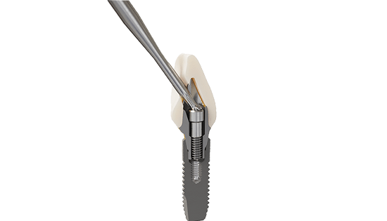
Typisk ved fortænder vil et skruehul til abutment være synligt, da skruens akse er i retning mod forsiden af kronen. Derfor har ASTRA Atlantis abutment udviklet et vinklet skrue system, således at skruehul kan skjules på bagsiden af kronen.
Referencer
Den publicerede litteratur understøtter brugen af Guided Implant til en forudsigelig og kontrolleret implantat kirurgi metode.
- Højere nøjagtighed sammenlignet med frihåndskirurgi (3–8)
- Sikker og forudsigelig operation kan anvendes alle steder i munden (2,4,12,18–28)
- Minimalt invasiv behandling er mulig (16,34,36)
- Reduceret tid i tandlægestolen kan opnås (37)
- Opretholdt patienttilfredshed ved årlige opfølgninger (38,39)
- Egbert N, Cagna DR, Ahuja S, Wicks RA. Accuracy and reliability of stitched cone-beam computed tomography images. Imaging Sci Dent. 2015 Mar;45(1):41-7 Abstract in Pubmed
- Stokbro K, Aagaard E, Torkov P, Bell RB, Thygesen T.
Virtual planning in orthognathic surgery. Int J Oral Maxillofac
Surg 2014;43(8):957-65. Abstract in PubMed
3. Vercruyssen M, Cox C, Coucke W, et al. A randomized
clinical trial comparing guided implant surgery (bone- or
mucosa-supported) with mental navigation or the use of
a pilot-drill template. J Clin Periodontol 2014;41(7):717-23.
Abstract in PubMed
4. Vercruyssen M, Coucke W, Naert I, et al. Depth and lateral
deviations in guided implant surgery: An rct comparing
guided surgery with mental navigation or the use of a pilotdrill template. Clin Oral Implants Res 2015;26(11):1315-20.
Abstract in PubMed
5. Shen P, Zhao J, Fan L, et al. Accuracy evaluation of
computer-designed surgical guide template in oral
implantology. J Craniomaxillofac Surg 2015;43(10):2189-94.
Abstract in PubMed
6. Arisan V, Karabuda CZ, Mumcu E, Ozdemir T.
Implant positioning errors in freehand and computer-aided
placement methods: A single-blind clinical comparative
study. Int J Oral Maxillofac Implants 2013;28(1):190-204.
Abstract in PubMed
7. Park C, Raigrodski AJ, Rosen J, Spiekerman C,
London RM. Accuracy of implant placement using precision
surgical guides with varying occlusogingival heights:
An in vitro study. J Prosthet Dent 2009;101(6):372-81.
Abstract in PubMed
8. Lin YK, Yau HT, Wang IC, Zheng C, Chung KH. A novel
dental implant guided surgery based on integration of
surgical template and augmented reality. Clin Implant Dent
Relat Res 2015;17(3):543-53. Abstract in PubMed
9. Edelmann AR, Hosseini B, Byrd WC, et al. Exploring
effectiveness of computer-aided planning in implant
positioning for a single immediate implant placement.
J Oral Implantol 2016;42(3):233-9. Abstract in PubMed
10. D’Haese J, De Bruyn H. Effect of smoking habits on
accuracy of implant placement using mucosally supported
stereolithographic surgical guides. Clin Implant Dent Relat
Res 2013;15(3):402-11. Abstract in PubMed
11. Cassetta M, Giansanti M, Di Mambro A, Stefanelli LV.
Accuracy of positioning of implants inserted using a
mucosa-supported stereolithographic surgical guide in
the edentulous maxilla and mandible. Int J Oral Maxillofac
Implants 2014;29(5):1071-8. Abstract in PubMed
12. Cassetta M, Di Mambro A, Giansanti M, Stefanelli LV,
Cavallini C. The intrinsic error of a stereolithographic surgical
template in implant guided surgery. Int J Oral Maxillofac
Surg 2013;42(2):264-75. Abstract in PubMed
13. Arisan V, Karabuda ZC, Ozdemir T. Accuracy of two
stereolithographic guide systems for computer-aided
implant placement: A computed tomography-based
clinical comparative study. J Periodontol 2010;81(1):43-51.
Abstract in PubMed
14. Testori T, Robiony M, Parenti A, et al. Evaluation of
accuracy and precision of a new guided surgery system:
A multicenter clinical study. Int J Periodontics Restorative
Dent 2014;34(suppl):s59-s69. Abstract in PubMed
15. Cassetta M, Di Mambro A, Giansanti M, Stefanelli LV,
Barbato E. Is it possible to improve the accuracy of implants
inserted with a stereolithographic surgical guide by reducing
the tolerance between mechanical components? Int J Oral
Maxillofac Surg 2013;42(7):887-90. Abstract in PubMed
16. Cassetta M, Di Mambro A, Di Giorgio G, Stefanelli LV,
Barbato E. The influence of the tolerance between
mechanical components on the accuracy of implants
inserted with a stereolithographic surgical guide:
A retrospective clinical study. Clin Implant Dent Relat Res
2015;17(3):580-8. Abstract in PubMed
17. Koop R, Vercruyssen M, Vermeulen K, Quirynen M.
Tolerance within the sleeve inserts of different surgical
guides for guided implant surgery. Clin Oral Implants Res
2013;24(6):630-4. Abstract in PubMed
18. Schneider D, Schober F, Grohmann P, Hammerle CH,
Jung RE. In-vitro evaluation of the tolerance of surgical
instruments in templates for computer-assisted guided
implantology produced by 3-d printing. Clin Oral Implants
Res 2015;26(3):320-5. Abstract in PubMed
19. D’haese J, Van De Velde T, Elaut L, De Bruyn H.
A prospective study on the accuracy of mucosally supported
stereolithographic surgical guides in fully edentulous
maxillae. Clin Implant Dent Relat Res 2012;14(2):293-303.
Abstract in PubMed
20. Van Assche N, Quirynen M. Tolerance within a
surgical guide. Clin Oral Implants Res 2010;21(4):455-58.
Abstract in PubMed
21. Al-Harbi SA, Sun AY. Implant placement accuracy
when using stereolithographic template as a surgical
guide: Preliminary results. Implant Dent 2009;18(1):46-56.
Abstract in PubMed
22. Arisan V, Karabuda ZC, Piskin B, Ozdemir T. Conventional
multi-slice computed tomography (ct) and cone-beam
ct (cbct) for computer-aided implant placement. Part
ii: Reliability of mucosa-supported stereolithographic
guides. Clin Implant Dent Relat Res 2013;15(6):907-17.
Abstract in PubMed
23. Cassetta M, Stefanelli LV, Giansanti M, Di Mambro A,
Calasso S. Accuracy of a computer-aided implant
surgical technique. Int J Periodontics Restorative Dent
2013;33(3):317-25. Abstract in PubMed
24. Cassetta M, Giansanti M, Di Mambro A, Calasso S,
Barbato E. Accuracy of two stereolithographic surgical
templates: A retrospective study. Clin Implant Dent Relat Res
2013;15(3):448-59. Abstract in PubMed
25. Cassetta M, Di Mambro A, Giansanti M, Stefanelli LV,
Barbato E. How does an error in positioning the template
affect the accuracy of implants inserted using a single fixed
mucosa-supported stereolithographic surgical guide? Int J
Oral Maxillofac Surg 2014;43(1):85-92. Abstract in PubMed
26. Stubinger S, Buitrago-Tellez C, Cantelmi G.
Deviations between placed and planned implant
positions: An accuracy pilot study of skeletally supported
stereolithographic surgical templates. Clin Implant Dent
Relat Res 2014;16(4):540-51. Abstract in PubMed
27. Valente F, Schiroli G, Sbrenna A. Accuracy of computeraided oral implant surgery: A clinical and radiographic
study. Int J Oral Maxillofac Implants 2009;24(2):234-42.
Abstract in PubMed
28. Van de Wiele G, Teughels W, Vercruyssen M, et al.
The accuracy of guided surgery via mucosa-supported
stereolithographic surgical templates in the hands of
surgeons with little experience. Clin Oral Implants Res
2014;E-pub Oct 16, doi:10.1111/clr.12494. Abstract in PubMed
29. Vercruyssen M, Cox C, Naert I, et al. Accuracy and
patient-centered outcome variables in guided implant
surgery: A rct comparing immediate with delayed
loading. Clin Oral Implants Res 2016;27(4):427-32.
Abstract in PubMed
30. Kang SH, Lee JW, Lim SH, Kim YH, Kim MK. Verification
of the usability of a navigation method in dental implant
surgery: In vitro comparison with the stereolithographic
surgical guide template method. J Craniomaxillofac Surg
2014;42(7):1530-5. Abstract in PubMed
31. Ruppin J, Popovic A, Strauss M, et al. Evaluation of
the accuracy of three different computer-aided surgery
systems in dental implantology: Optical tracking vs.
Stereolithographic splint systems. Clin Oral Implants Res
2008;19(7):709-16. Abstract in PubMed
32. Sarment DP, Sukovic P, Clinthorne N. Accuracy of implant
placement with a stereolithographic surgical guide. Int J Oral
Maxillofac Implants 2003;18(4):571-7. Abstract in PubMed
33. Somogyi-Ganss E, Holmes HI, Jokstad A. Accuracy of
a novel prototype dynamic computer-assisted surgery
system. Clin Oral Implants Res 2015;26(8):882-90.
Abstract in PubMed
34. Abboud M, Wahl G, Guirado JL, Orentlicher G.
Application and success of two stereolithographic surgical
guide systems for implant placement with immediate
loading. Int J Oral Maxillofac Implants 2012;27(3):634-43.
Abstract in PubMed
35. Aboul-Hosn Centenero S, Hernandez-Alfaro F.
3d planning in orthognathic surgery: Cad/cam surgical
splints and prediction of the soft and hard tissues results
– our experience in 16 cases. J Craniomaxillofac Surg
2012;40(2):162-8. Abstract in PubMed
36. Arisan V, Bolukbasi N, Oksuz L. Computer-assisted
flapless implant placement reduces the incidence of surgeryrelated bacteremia. Clin Oral Investig 2013;17(9):1985-93.
Abstract in PubMed
37. Arisan V, Karabuda CZ, Özdemir T. Implant surgery
using bone- and mucosa-supported stereolithographic
guides in totally edentulous jaws: Surgical and postoperative outcomes of computer-aided vs. Standard
techniques. Clin Oral Implants Res 2010;21(9):980-88.
Abstract in PubMed
38. Van de Velde T, Sennerby L, De Bruyn H. The clinical and
radiographic outcome of implants placed in the posterior
maxilla with a guided flapless approach and immediately
restored with a provisional rehabilitation: A randomized
clinical trial. Clin Oral Implants Res 2010;21(11):1223-33.
Abstract in PubMed
39. Vercruyssen M, van de Wiele G, Teughels W, et al.
Implant- and patient-centred outcomes of guided surgery,
a 1-year follow-up: An rct comparing guided surgery
with conventional implant placement. J Clin Periodontol
2014;41(12):1154-60. Abstract in PubMed
